
Your Knee
Your knee is made up of bone, cartilage, meniscus, multiple ligaments, tendons, bursa and muscles. These structures can be affected by an injury, a degenerative process or other rare diseases. Problems with the knee can manifest in numerous ways and may include pain, swelling, instability or locking.
Dr Hirner offers comprehensive management of all knee conditions. If surgery is required he may use a number of techniques which include keyhole (arthroscopy), computer assisted and conventional surgery.
Knee Anatomy
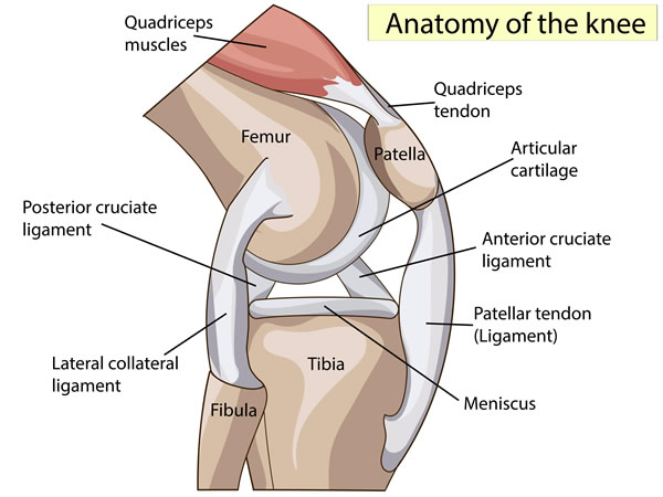
The adjacent diagram displays the anatomical structures of the knee. The bones, femur, tibia and patella are lined by articular cartilage which allows the boney surfaces to glide smoothly. Between the bones is the meniscus which helps with knee stability and also produces a shock absorbing affect. The multiple ligaments ACL (anterior cruciate ligament), PCL (posterior cruciate ligament) MCL (medial collateral ligament) and LCL (lateral collateral ligament) generally offer a stabilizing effect. The muscles surrounding the knee (quadriceps, hamstrings and others) are powerful mobilizers of the joint.
Knee Conditions
Dr. Hirner treats all knee problems and this is a list of some of the most common conditions (Click on the link for more information)
- Anterior cruciate ligament (ACL) tears
- Posterior cruciate ligament (PCL) tears
- Medial collateral ligament or lateral collateral ligament (MCL or LCL) tears
- Patellofemoral instability (knee cap dislocation)
- Meniscus tears - video
- Articular cartilage injury
- Osteochondral injury
- Osteonecrosis (SONK)
- Osteoarthritis
- Patella arthritis
- Pre patella bursitis
- Quads and patella tendon rupture
- Patellofemoral pain syndrome
- Osteotomy around the knee
- Unicompartment Knee Replacement
- Viscosupplementation in the Knee
- Platelet rich plasma
- Pigmented villanodular synovitis
- Osteochondritis dessicans
- Adolescent anterior knee pain
- Common knee injuries
- Discoid meniscus
- Patellofemoral arthritis
Rehabilitation Protocols
(Click on the link for protocols to follow)
- ACL rupture
- PCL reconstruction
- Multi-ligament reconstruction
- Meniscal tear
- Meniscal repair
- Meniscal posterior root repair
- Patella instability (MPFL reconstruction)
- High tibial osteotomy
- Total knee replacement
Disclaimer
Northland Orthopaedic Center Management LTD is a shareholder in the Northland MRI scan. Unfortunately this is the only private MRI scanner in Northland. If however you would prefer to be referred to an alternative provider in Auckland please inform Dr Hirner and his staff will make an appropriate referral.
Kensington Hospital has received funding from Zimmer Biomet and Lima for the Northland Orthopaedic Training Fellowship in shoulder & knee surgery. Dr Hirner has lectured for Smith & Nephew, Arthrex, Zimmer Biomet.
Dr Hirner is a trustee of the Northland Orthopaedic Charitable Research Trust.
Southern Cross Northland Surgical Centre, 15 Kensington Ave, Whangarei, 0112
P: 09 437 9027
![]()

© Marc Hirner 2026. All rights reserved. Energise Website Design